
Phase Contrast in Tomography - Pellets
Case Study: Visualization of Pellets - Materials and Porosity
In collaboration with Danish company Biomar, DTI has investigated batches of experimental fish feed pellets. Biomar is interested in how the experimental diets affect the digestion process of fish, which is why they see a great potential in analyzing their material by non-destructive 3D imaging.
From Laboratory Micro-CT to HiP Synchrotron Tomography
- To begin with, the pellets, less than a millimeter in diameter, were analyzed in our laboratory micro-CT scanner (high resolution 3D X-ray tomography). The Micro-CT scanner can produce virtual cross sections images of materials at high resolution (down to 1 µm voxel size). These images were a valuable resource to compare the internal structure of different pellet samples, and the technique is non-destructive and therefore visualizes for example internal porosities very well.
- Seeing the potential to quantify data, the samples were then also analyzed by phase contrast synchrotron tomography (HiP-CT) at the European Synchrotron Radiation Facility (ESRF) in Grenoble, France. The synchrotron source itself will give a much better signal-to-noise ratio than a laboratory Micro-CT, but as an added benefit, the phase contrast will greatly increase the contrast in for example organic material, as can be clearly seen in the comparison below.
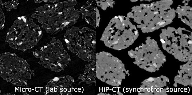
Comparison of a cross section from laboratory micro-CT (left) and synchrotron phase contrast HiP-CT (right)
The overarching gain to include this top-of-the-art technology is to continue improving what we offer to the aquaculture industry in form of a feed. In other words, each pellet we produced is based on solid science, and this exciting collaboration provided us with the right tools.
Pedro Gómez, Senior Scientist, Biomar Denmark
Quantification of 3D tomography data
With the enhanced image quality possible with the synchrotron source, reliable quantification of materials and porosity could be performed (quantification not published). In the visualization below, the internal porosity of a single pellet is shown. It could be determined that almost all porosity connected to the surface of the pellet - only a small fraction was closed in by material. Characteristics like these influence how the pellets function and how they dissolve.
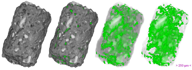
3D visualization of porosity in a single pellet (HiP synchrotron tomography)
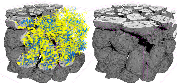
3D visualization of internal material distribution (HiP synchrotron tomography)
Danish Technological Institute's role as a mediator
At DTI, we can help you obtain access to exciting techniques like HiP-CT. We can help you with the whole process from experimental design, to data collection, and data analysis. Have you ever thought about using big science to push your research forward?
Get in touch with us!
- Olivia Aalling-Frederiksen, Specialist (ofr@dti.dk / +45 7220 3042)
____________________
ACKNOWLEDGEMENT:
This study was supported by a grant from Danish Technological Institutes performance contract 2021-2024, entered with the Danish Agency for Higher Education and Science, under The Ministry of Higher Education and Science Denmark. Collaborators at Biomar and ESRF are greatly acknowledged.