Imaging of biological and medico materials - Synchrotron Tomography
Synchrotron Tomography - Non-Destructive Imaging of Low-Density Materials
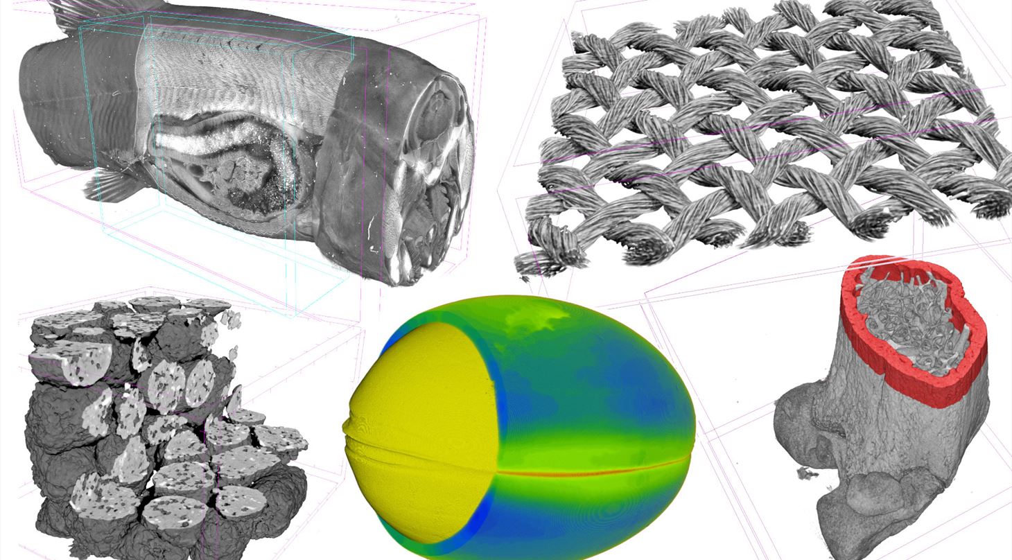
Low density materials, including biological samples and materials composed primarily of organic compounds, can be hard to image in 3D because they have a low X-ray absorption. X-ray absorption increases with an element’s atomic number, and therefore materials made of carbon (atomic number 6) absorb only a small amount of the irradiation, thus giving very poor contrast in imaging.
With the addition of phase contrast tomography, this is all about to change. Using phase contrast, even biological samples can be studied with high resolution and contrast - without staining or similar invasive sample preparation.
Low density materials can be hard to image because they have a low X-ray contrast. With phase contrast, we see a game-changer for our customers in the food- and medico industries
- Susan Cooper, Team Manager, DTI
HiP-CT (Hierarchical Phase-Contrast Tomography)
One of the common ways to image biological materials has been to use a contrast agent, where a light material is treated with heavy elements that absorb into the material to increase the contrast. Another way to image materials made of light elements is by measuring samples using phase contrast, where changes in material density determine the image contrast instead of elemental identity. In addition, synchrotron sources can be used instead of lab X-ray sources so we get more signal.
This type of analysis can be used on anything made of light elements, including:
- Biological samples
- Food
- Composites
- Pharmaceuticals
Danish Technological Institute's Role as a Mediator
At DTI, we can help you obtain access to exciting techniques like HiP-CT. We can help you with the whole process from experimental design, to data collection, and data analysis. Have you ever thought about using big science to push your research forward? What do you want to see in detail?
Get in Touch With Us!
- Susan Rudd Cooper (srco@dti.dk / +45 7220 1754)