Visualization of Soft Tissue in Small Fish - Phase Contrast CT
Case Study: Visualization of Soft Tissue in Small Fish
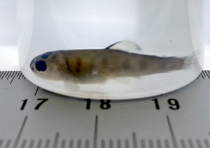
In collaboration with the Danish company Biomar, DTI has investigated batches of small fish after being fed with different experimental diets. Biomar is interested in how the experimental diets affect the digestion process of the fish, which is why they saw a great potential in non-destructive 3D imaging as a complement to dissection and histological analysis.
Our collaboration with DTI has tremendously aid on expanding our research tools to increase our knowledge on fish physiology. The overarching gain to include this top-of-the-art technology is to continue improving what we offer to the aquaculture industry in form of a feed. In other words, each pellet we produced is based on solid science, and this exciting collaboration provided us with the right tools.
Pedro Gómez, Senior Scientist, Biomar Denmark
From Laboratory Micro-CT to Phase Contrast Synchrotron Tomography
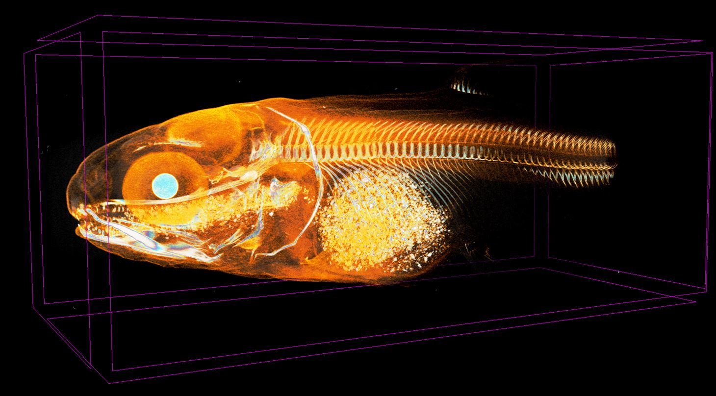
 3D scans were first performed on a laboratory micro-CT available at DTI, a procedure which, for this type of specimen, includes heavy metal staining to increase X-ray absorption in the low density tissue. A collaboration with the European Synchrotron Radiation Facility (ESRF) in France was then initiated, which led to the project continuing with their new state-of-the-art imaging technique, HiP-CT (Hierarchical Phase-Contrast Tomography). Data quality was drastically improved with the introduction of phase contrast-CT, and heavy metal staining was no longer necessary. The much-improved level of detail and contrast seen in the soft tissues of the fish was made possible by the phase contrast and high flux achieved at the synchrotron facility.
This is the most jaw-dropping data I've ever worked on! Many of our customers come from the food or pharmaceutical industry, and I can easily see how they could benefit from this type of analysis.
Erik Wisaeus, Business Manager, DTI
Ground-Breaking Phase Contrast Tomography
The data from scanning of these small fish has excited our imaging expert Erik, who says, “I want to make it abundantly clear that this is The Most Jaw-Dropping Data I’ve ever worked on. While we got good and useful data from our micro-CT analysis, phase contrast tomography at the synchrotron facility really set the bar higher with the extreme level of detail we now can see. This assignment has really opened my eyes to the possibilities made available by collaborating with the big science facilties. Many of our customers come from the food or pharmaceutical industry, and I can easily see how they could benefit from this type of analysis.”
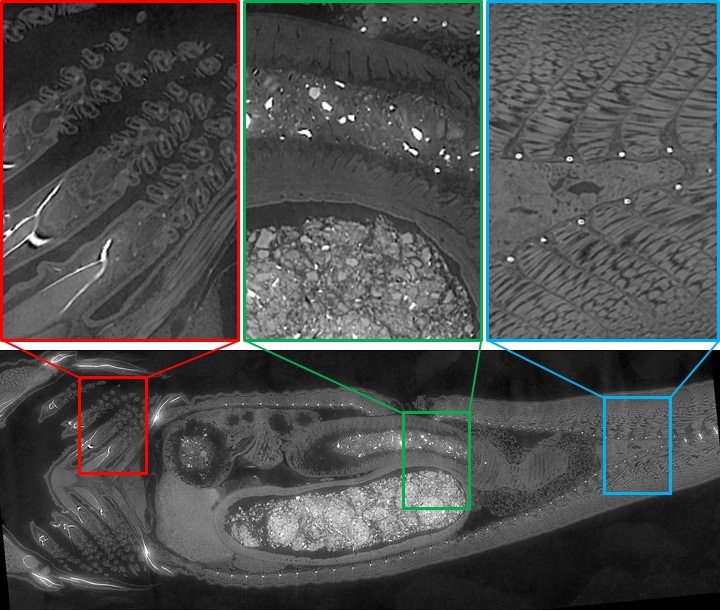
The collage below shows example detail images from a single virtual cross section of an unstained small fish of ca 25 mm length. The insets show an incredible level of detail, here exemplified in a region of the gills, the stomach/intestine, and an area near the rectum. The collected data makes it possible to study how different experimental diets are processed by fish. Apart from a series of cross section images, a variety of 3D visualizations were produced to be able to compare the various specimens.
Phase contrast-CT is a new tool to see unprecedented detail in soft tissues and other organic material, and will revolutionize how we analyze food products, medical products and tissue for research and product development. The highest resolution that can be reached using phase contrast-CT at a synchrotron facility is 100 times that of a medical CT scanner, and contrary to regular CT scanning, good contrast can now be achieved also in soft tissue and other low-density materials.
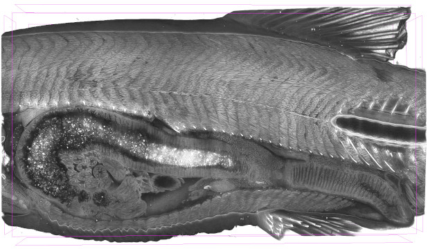
3D Visualization Makes Huge Datasets Easy to Navigate
Tomography datasets typically contain a huge amont of information. At DTI, we are experienced with both visualization and quantification of tomographical data. We can produce videos that makes it easy to understand the sample structure, and to compare different samples between each other.
Looking at the fish data makes you feel like you are getting to experience a new part of the world that has, until recently, not been possible to see!
Susan Cooper, Team Manager, DTI
Danish Technological Institute's Role as a Mediator
At DTI, we can help you obtain access to exciting techniques like phase contrast-CT. We can help you with the whole process from experimental design, to data collection, and data analysis. Have you ever thought about using big science to push your research forward?
Get in Touch With Us!
- Susan Rudd Cooper (srco@dti.dk / +45 7220 1754)
____________________
ACKNOWLEDGEMENT:
This study was supported by a grant from Danish Technological Institutes performance contract 2021-2024, entered with the Danish Agency for Higher Education and Science, under The Ministry of Higher Education and Science Denmark. Collaborators at Biomar and ESRF are greatly acknowledged.