
High-energy X-ray radiation reveals hidden weaknesses in high-strength bolts
High-strength bolts can play a significant role in future wind turbine design. They allow for slender structures, lower material consumption, and better fatigue properties – but only if the quality is in order. When a bolt failed mechanical testing in a development project, the Danish Technological Institute resorted to an advanced characterization method to learn more: High-energy X-ray diffraction.
High-strength bolts – heat-treated steel bolts – hold great potential in, for example, the wind industry. But when strength is increased, the demands on process control also grow, as small variations in heat treatment and microstructure can have a major impact on the bolt's mechanical properties.
The metallurgical specialists at the Danish Technological Institute experienced this during the characterization of a heat-treated bolt that did not meet the specified mechanical requirements. Using high-energy X-ray diffraction at the DESY synchrotron (Deutsches Elektronen-Synchrotron) in Hamburg, a cross-section of the bolt was analyzed with a level of detail that goes far beyond classical metallography.
From mechanical testing to microstructural explanation
The original task involved the development of large-scale high-strength bolts – a product currently not available on the market, but with great potential for design optimization and cost savings. The client was a wind turbine manufacturer who wanted to be certain of the bolts' quality before they could be implemented in the company's designs. The bolts were to be tested for tensile strength, fatigue properties, and the risk of hydrogen embrittlement, among other things – classic parameters when components must be qualified for demanding industrial applications.
Subsequently, the Danish Technological Institute proceeded with an advanced microstructural analysis of a bolt that failed the mechanical tests. This was done as part of the performance contract 'Advanced characterization of products and processes at large-scale facilities', supported by the Danish Agency for Higher Education and Science. The goal was to investigate whether the explanation for the bolt's failure could be found in the internal structure of the material.
High-strength bolts are typically manufactured by heat-treating carbon steel, and the heat treatment is precisely what is critical. Among other things, it controls the formation and distribution of crystalline phases in the steel – including ferrite and cementite. If the phase distribution is not homogeneous, it can create local variations in strength and toughness
- Nils Lau Nyborg Broge, Danish Technological Institute
Synchrotron measurement with 1.3 million data points
At DESY, a cross-section of a bolt was scanned using high-energy X-ray radiation – also known as X-ray diffraction imaging. This is an effective, non-destructive analysis method used to map the structure of crystalline materials. It works by bombarding a material with X-rays and measuring how the rays are scattered by the material's electrons. This yields a unique fingerprint of the substance's internal structure.
Instead of simply forming an image, the method measures X-ray diffraction at every single point. This means that not only do you see the structure – you also obtain information about which crystallographic phases are located where in the material.
The measurement resulted in 1.3 million diffractograms and was completed in about 45 minutes. Each measurement point had a spatial resolution of approximately 2 µm x 20 µm, providing an extremely detailed phase map of the bolt's cross-section.
It is precisely the combination of speed, resolution, and data quality that makes this technology interesting. In principle, much of this information can be obtained using other laboratory techniques, but rarely on the same scale and never in such a short amount of time.
Cementite gradient revealed inhomogeneous phase distribution
The analysis showed that the bolt primarily consisted of the crystalline phase ferrite, which is expected for steel, as well as smaller amounts of cementite (Fe3C). Additionally, the iron oxide phase magnetite (Fe3O4), also known as black oxide, was identified near the surface, which is consistent with the manufacturing process.
However, the crucial finding was the distribution of cementite. The phase was not homogeneously distributed across the cross-section but showed a clear overrepresentation near the outer edge of the bolt. There was both a slight gradient from the center towards the surface and an actual precipitation close to the edge.
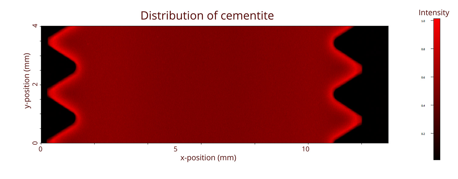
– This suggests that the heat treatment was not sufficiently controlled. An uneven distribution of carbon and cementite can lead to localized differences in mechanical properties – and thus be a likely explanation for why the bolt did not achieve the expected strength, says Nils Lau Nyborg Broge, specialist in corrosion and metallurgy at the Danish Technological Institute.
A similar analysis can also be expected to provide a wealth of information about other types of components and materials, particularly if they are composed of multiple phases or subcomponents.
A powerful tool for quality and process development
For the industry, the value does not lie solely in explaining a single failure. The method makes it possible to quickly compare good and defective parts, document phase distributions and homogeneity, and link microstructure to mechanical properties. Thus, high-energy X-ray diffraction allows for deep insight into the internal structure of components, which is crucial for the efficient development of better products.
Compared to traditional metallographic characterization, synchrotron-based X-ray diffraction can deliver much larger amounts of data in less time and with less manual laboratory work. The results can be quantitatively analyzed to a high degree of detail, but also quickly and directly visualized as phase maps, where qualitative characteristics become apparent.
Advanced characterization closer to the industry
This case shows that synchrotron technology is not reserved solely for basic research. When the samples are relevant and the analysis is correctly designed, high-energy X-ray measurements can provide industrial value on a week-to-week basis.
For companies working with critical metal components, the perspective is clear: they can gain access to a detailed insight into the material's internal state – quickly, relatively cost-effectively, and with a resolution that can reveal failure mechanisms before they lead to breakdowns. At the Danish Technological Institute, we stand ready to help, so please contact us if you need characterization using high-energy X-ray radiation.
Specifications
| Sample dimensions | 12 mm x 4 mm x 3 mm |
| Pixel size | 2 µm x 20 µm |
| Total diffractograms measured | 1.300.000 |
| Measurement rate | 500 Hz |
| Beamline | P07, DESY |
| X-ray energy | 73.3 keV |
| Detector | Dectris Eiger CdTe 4m |